
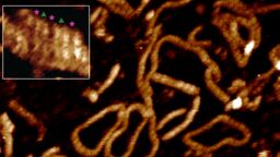
Scientists at the University of Sheffield, in collaboration with the University of York, have achieved a landmark breakthrough in molecular biology by directly visualizing two DNA double helices locking together for the first time. This discovery resolves a mechanism that has puzzled researchers for more than twenty years: how DNA molecules overcome their identical negative charges to pair up inside living cells.
Breaking Through the Charge Barrier
DNA carries a strong negative charge along its phosphate backbone. According to basic electrostatic principles, like charges repel. Yet inside every cell, DNA strands must recognize and pair with each other to carry out essential functions including genetic recombination, gene silencing, and chromosome packaging. For decades, the “DNA zipper” model proposed by Professor Alexey Kornyshev at Imperial College London offered a theoretical explanation, but direct experimental evidence remained elusive.
The research team, led by Dr. Thomas Catley from the School of Chemical Materials and Biological Engineering at Sheffield and Professor Agnes Noy from the School of Physics, Engineering and Technology at York, employed high-powered atomic force microscopy (AFM) to scan DNA samples and build topographical maps at the molecular level. What they observed confirmed the theoretical predictions: short DNA fragments matching up with exact precision, groove for groove, like interlocking spiral staircases.
The Molecular Bridge: Divalent Metal Ions
Advanced computer simulations, led by Dr. Victor Velasco-Berrelleza from Sheffield’s School of Mathematical and Physical Sciences, revealed the mechanism behind this pairing. Positively charged metal ions—specifically divalent cations including nickel, magnesium, and calcium—act as tiny molecular bridges. These ions nestle inside the grooves of the DNA double helix, with each ion using its two positive charges to hold both strands simultaneously across the gap.
“While microscopy can show us what happens, it’s the simulations that allow us to uncover the molecular mechanism behind it,” explained Dr. Velasco-Berrelleza. “This mechanism could be another tool in the DNA regulatory toolkit, where some DNA sequences may not encode proteins but could instead influence where DNA molecules interact with each other, helping the genome form higher-order structures such as chromosomes.”
Sequence-Specific Pairing Hotspots
One of the most significant findings is that DNA pairing is not uniform across the genome. Certain DNA sequences form much stronger contacts than others, creating specific hotspots where two helices are particularly likely to align. This sequence dependence suggests that the genome may have evolved to exploit these preferential pairing regions for regulatory purposes.
Professor Agnes Noy emphasized the implications: “This discovery could help researchers identify regions of the genome specially involved in DNA pairing. These regions may become particularly important when mutations disrupt normal cellular processes and contribute to cancer.”
Implications for Cancer Research and Biotechnology
The ability to identify and characterize DNA pairing hotspots opens new avenues for cancer research. When mutations occur in these critical regions, they may disrupt normal DNA-DNA interactions, leading to genomic instability—a hallmark of cancer. Understanding these mechanisms could lead to new diagnostic markers or therapeutic targets.
Beyond medical applications, the findings have significant implications for DNA nanotechnology and biotechnology. Dr. Catley noted: “Hopefully, these programmable interactions could eventually help engineers design custom DNA structures for future biotechnology, such as DNA origami and shed light on how DNA is actually packaged inside cells.”
Curiosity-Driven Research with Societal Impact
The study, published in Nucleic Acids Research under the title “Imaging and mechanism of DNA–DNA recognition mediated by divalent ions,” exemplifies how fundamental, curiosity-driven research can align with wider societal priorities. The project brought together expertise in advanced imaging, computational modeling, and molecular biology across two leading UK universities.
The University of Sheffield, a Russell Group institution ranked among the world’s top 100 universities, continues to demonstrate its commitment to pioneering research that addresses fundamental scientific questions while creating pathways for practical applications in medicine and technology.
Next Steps in DNA Interaction Research
With the mechanism now visualized and understood at the atomic level, several research directions emerge. The team plans to investigate how proteins and other cellular factors modulate these DNA-DNA interactions in vivo. Additionally, mapping pairing hotspots across the human genome could reveal new regulatory elements and provide insights into the three-dimensional organization of chromosomes.
For researchers and students interested in pursuing work at the intersection of biophysics, molecular biology, and computational modeling, this breakthrough highlights the power of interdisciplinary approaches. The combination of cutting-edge microscopy with atomistic simulations proved essential—neither technique alone could have revealed the complete picture.
Explore postgraduate research opportunities in molecular biology and biophysics at the University of Sheffield to contribute to the next generation of discoveries in DNA science.
Understanding the Experimental Approach
Atomic Force Microscopy: Seeing the Invisible
Atomic force microscopy operates by scanning a sharp tip across a sample surface, measuring forces between the tip and the sample to construct a topographical map with nanometer resolution. Unlike electron microscopy, AFM can operate in liquid environments, making it suitable for imaging biological molecules in near-physiological conditions. The Sheffield-York team optimized this technique to capture the transient DNA-DNA interactions that occur in solution.
Molecular Dynamics Simulations: The Computational MicroscopeComplementing the experimental imaging, molecular dynamics simulations tracked the movement of individual atoms and ions over time. These computational models, requiring substantial computing power, revealed the dynamic behavior of divalent cations in the DNA grooves—information impossible to obtain from static images alone. The simulations confirmed that each divalent ion coordinates with both DNA strands simultaneously, forming a stable bridge that overcomes electrostatic repulsion.
Historical Context: The DNA Zipper Model
The “DNA zipper” model, first proposed over two decades ago by Professor Alexey Kornyshev and collaborators, suggested that surrounding salt ions create alternating charge patterns along the DNA helix, allowing molecules to line up like interlocking spiral staircases. The model was elegant but remained theoretical until now. The Sheffield-York collaboration provides the first direct experimental validation, transforming a theoretical framework into an observed biological mechanism.
This validation process illustrates the scientific method at work: a theoretical prediction guides experimental design, advanced instrumentation enables observation, and computational modeling explains the underlying physics. Each component was essential, and the collaboration across disciplines and institutions made the breakthrough possible.
Broader Implications for Genome Biology
DNA-DNA interactions extend beyond the specific pairing observed in this study. The genome’s three-dimensional organization—how meters of DNA fit into a micron-scale nucleus while remaining accessible for transcription, replication, and repair—depends on a hierarchy of interactions. DNA-DNA pairing mediated by divalent cations represents a fundamental layer of this organization.
Understanding these interactions may also shed light on phase separation phenomena in the nucleus, where DNA and proteins condense into distinct compartments. The sequence-specific pairing hotspots identified in this study could serve as nucleation points for such higher-order structures.
Schedule a consultation with our research admissions team to discuss PhD projects in genome biology and molecular biophysics.
Training the Next Generation of Interdisciplinary Scientists
This research highlights the growing importance of interdisciplinary training in modern biology. The first author, Dr. Victor Velasco-Berrelleza, bridges mathematics, physics, and biology—exemplifying the hybrid skill set increasingly required for breakthrough discoveries. The University of Sheffield’s cross-faculty research centers are designed to foster exactly this type of collaborative, interdisciplinary work.
For prospective students, this means opportunities to work at the cutting edge of multiple fields simultaneously. Programs in biological physics, computational biology, and molecular engineering prepare graduates for research careers that transcend traditional departmental boundaries.
From Fundamental Discovery to Clinical Application
The path from fundamental discovery to clinical application is rarely direct, but understanding DNA-DNA recognition mechanisms creates multiple potential translational pathways. Cancer genomics could benefit from identifying pairing hotspots as potential biomarkers for genomic instability. Gene therapy approaches might leverage programmable DNA-DNA interactions for targeted genome editing. DNA nanotechnology applications, from drug delivery vehicles to molecular computing, gain a new design principle.
The research team’s emphasis on “programmable interactions” suggests a future where DNA serves not just as a passive information storage molecule but as an active, engineerable component in synthetic biology systems. The ability to predict and control DNA-DNA pairing based on sequence and ionic conditions provides a new toolkit for bioengineers.
Discover how our MSc programs in Molecular Biology and Biotechnology prepare students for careers at the forefront of genetic engineering and genomic medicine.
The Role of Metal Ions in Cellular Biology
Beyond DNA pairing, the finding that divalent cations serve as molecular bridges has broader implications for cellular biology. Magnesium and calcium ions are ubiquitous in cells, with tightly regulated concentrations. Their role in mediating DNA-DNA interactions adds another layer to our understanding of how ionic homeostasis affects genome function. Disruptions in metal ion homeostasis—observed in various diseases—may have previously unrecognized effects on DNA organization and gene regulation.
This connection between inorganic ion biology and genome architecture represents an emerging research frontier. The Sheffield-York team’s methodology—combining high-resolution imaging with atomistic simulations—provides a template for investigating other ion-mediated biomolecular interactions.
Collaborative Research Excellence in the UK
This breakthrough emerged from collaboration between two leading UK universities, each contributing complementary expertise. The University of Sheffield’s strength in advanced materials characterization and the University of York’s excellence in computational physics created a partnership greater than the sum of its parts. Such collaborations are increasingly essential for tackling complex biological questions that span multiple scales—from atomic interactions to cellular function.
The UK’s research ecosystem, with its concentration of world-class universities and supportive funding structures, continues to produce globally significant discoveries. The publication in Nucleic Acids Research, a leading journal in the field, ensures this work reaches the international scientific community and stimulates further research worldwide.
Learn more about collaborative research opportunities and international partnerships at the University of Sheffield’s research centers.
Looking Ahead: Unanswered Questions
While this study resolves a decades-old mystery, it also raises new questions. How do DNA-binding proteins compete with or cooperate with cation-mediated DNA-DNA interactions? Do pairing hotspots correspond to known regulatory elements or reveal novel ones? How do epigenetic modifications affect pairing propensity? What role do these interactions play in meiotic recombination versus somatic genome organization?
The research team is already pursuing several of these questions. The methodology they developed—correlative AFM and simulation—can now be applied to more complex systems, including protein-DNA complexes and nucleosome arrays. Each answer will likely reveal new layers of complexity in how the genome organizes and regulates itself.
Conclusion
The direct visualization of DNA strands zipping together represents a milestone in molecular biology. By solving the long-standing puzzle of how DNA overcomes electrostatic repulsion to pair specifically, the University of Sheffield and University of York researchers have opened new avenues for understanding genome organization, cancer biology, and DNA nanotechnology. The discovery validates the power of interdisciplinary approaches combining advanced imaging, computational modeling, and molecular biology.
For the scientific community, this work provides a foundation for investigating DNA-DNA interactions in their full biological context. For students and early-career researchers, it demonstrates the excitement and impact of working at disciplinary boundaries. For society, it exemplifies how fundamental research—driven by curiosity about nature’s basic mechanisms—creates knowledge that ultimately enables medical advances and technological innovation.
The study, “Imaging and mechanism of DNA–DNA recognition mediated by divalent ions,” is available in Nucleic Acids Research. As research continues to build on this foundation, the implications for cancer research, biotechnology, and our understanding of life’s molecular machinery will continue to unfold.
Have questions about this research or related study programs? Write to us for more information.
Share your thoughts on this breakthrough in the comments below and explore our related articles on genomic medicine and molecular biophysics.